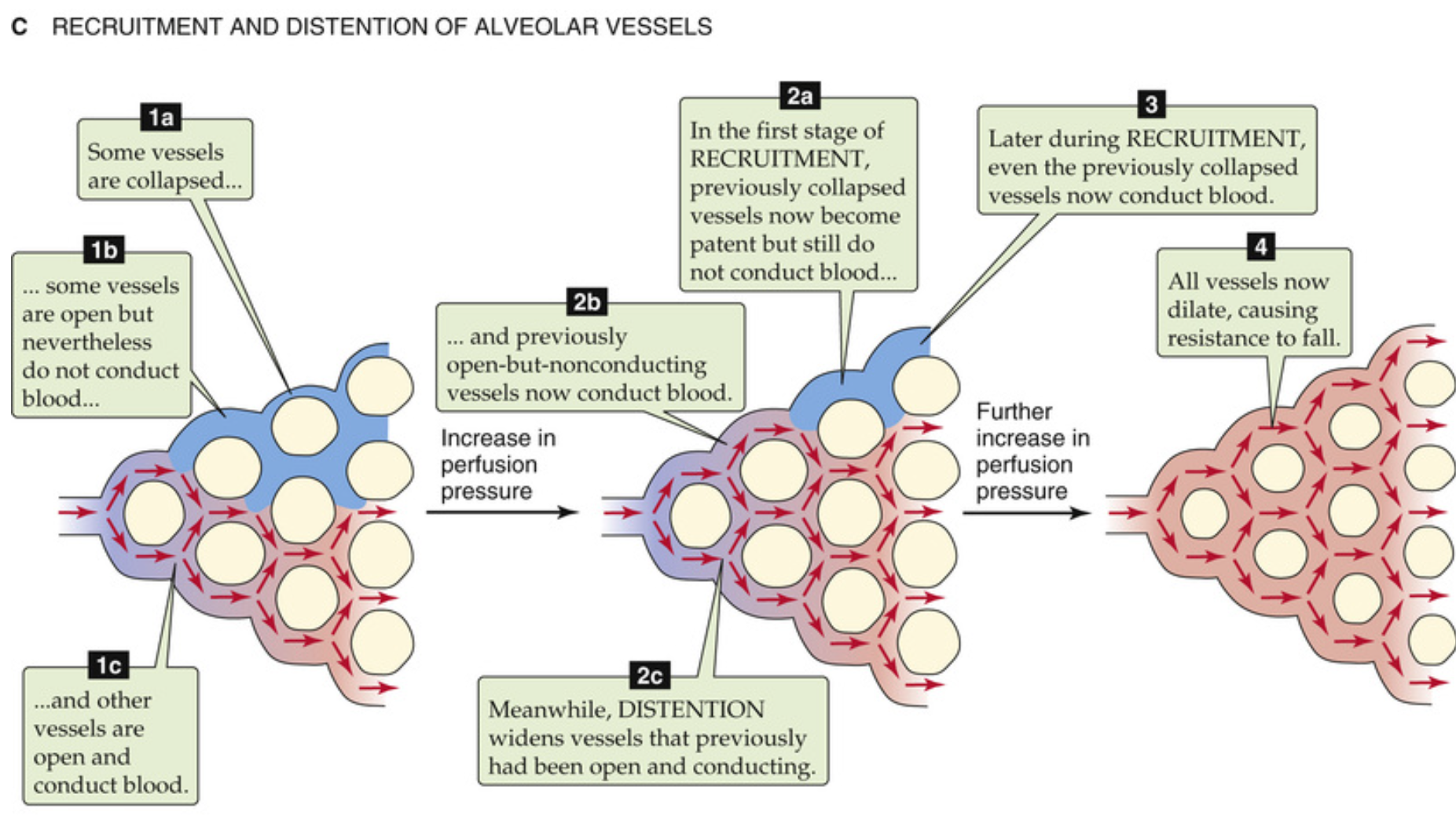
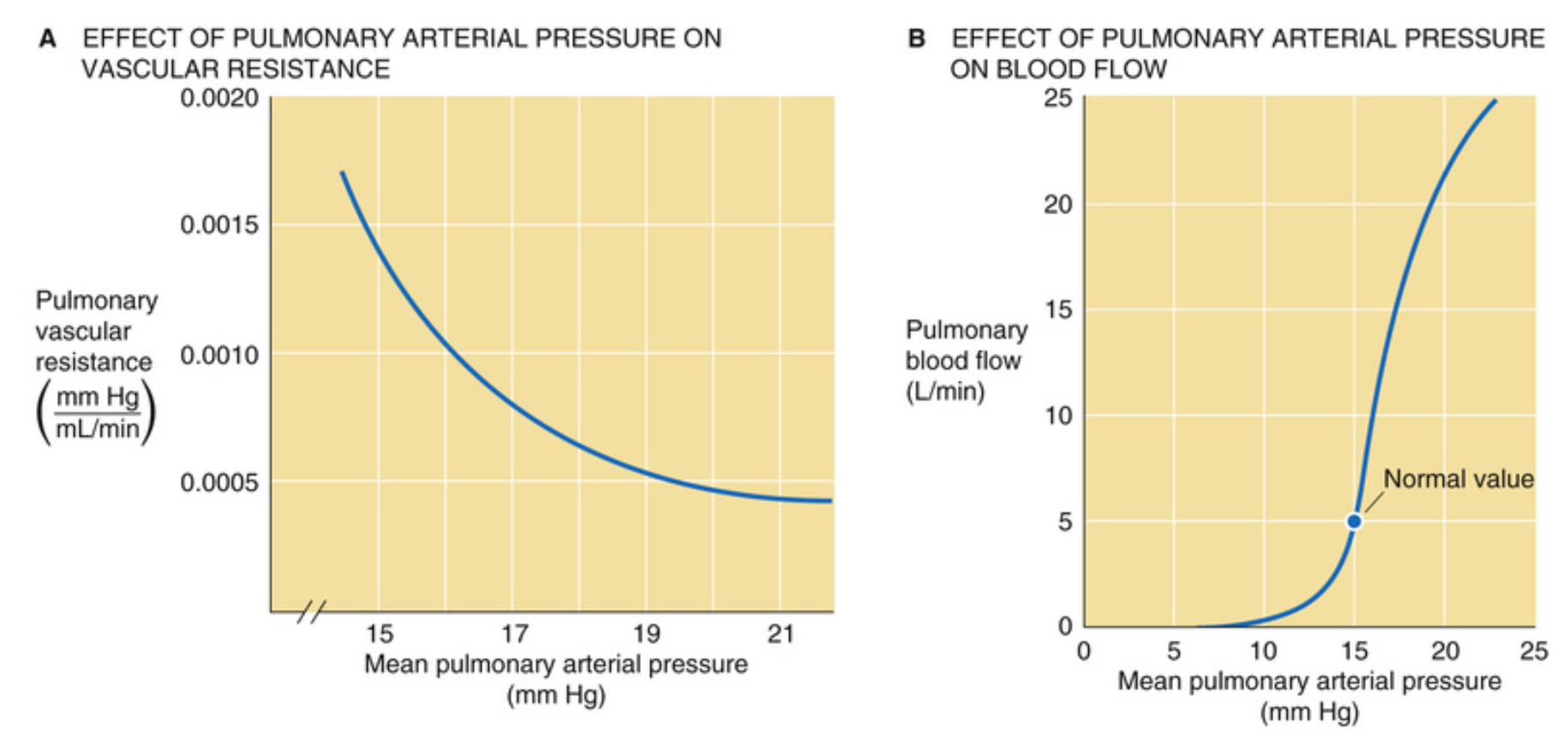
Created by Jiří Kofránek
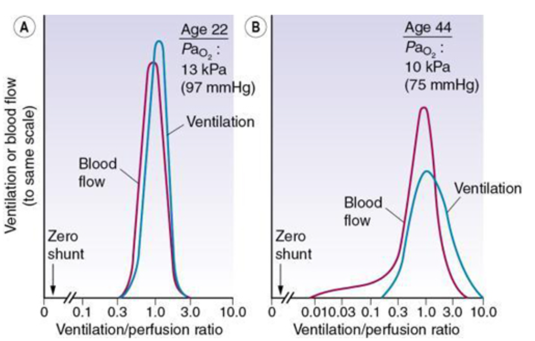
(A) - A male aged 22 years (B) The wider spread of ratios in a male aged 44 years.
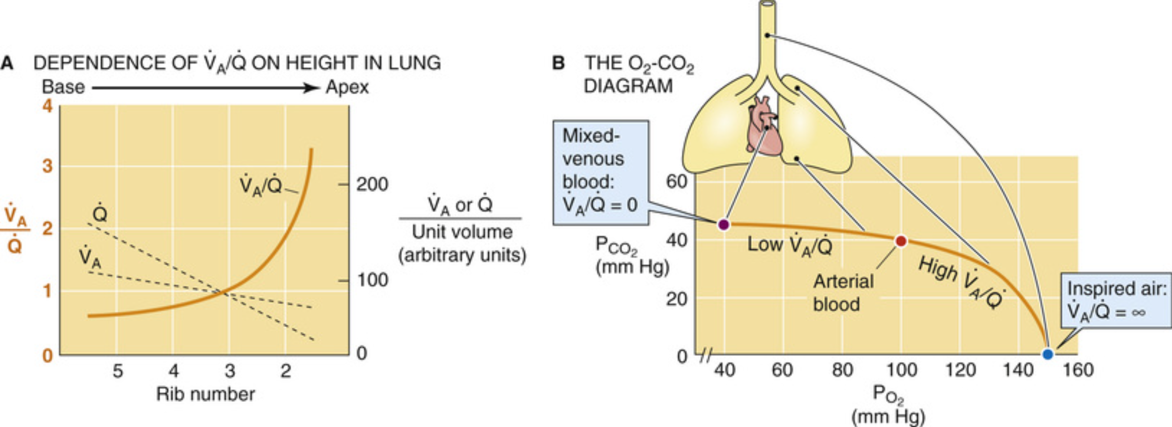
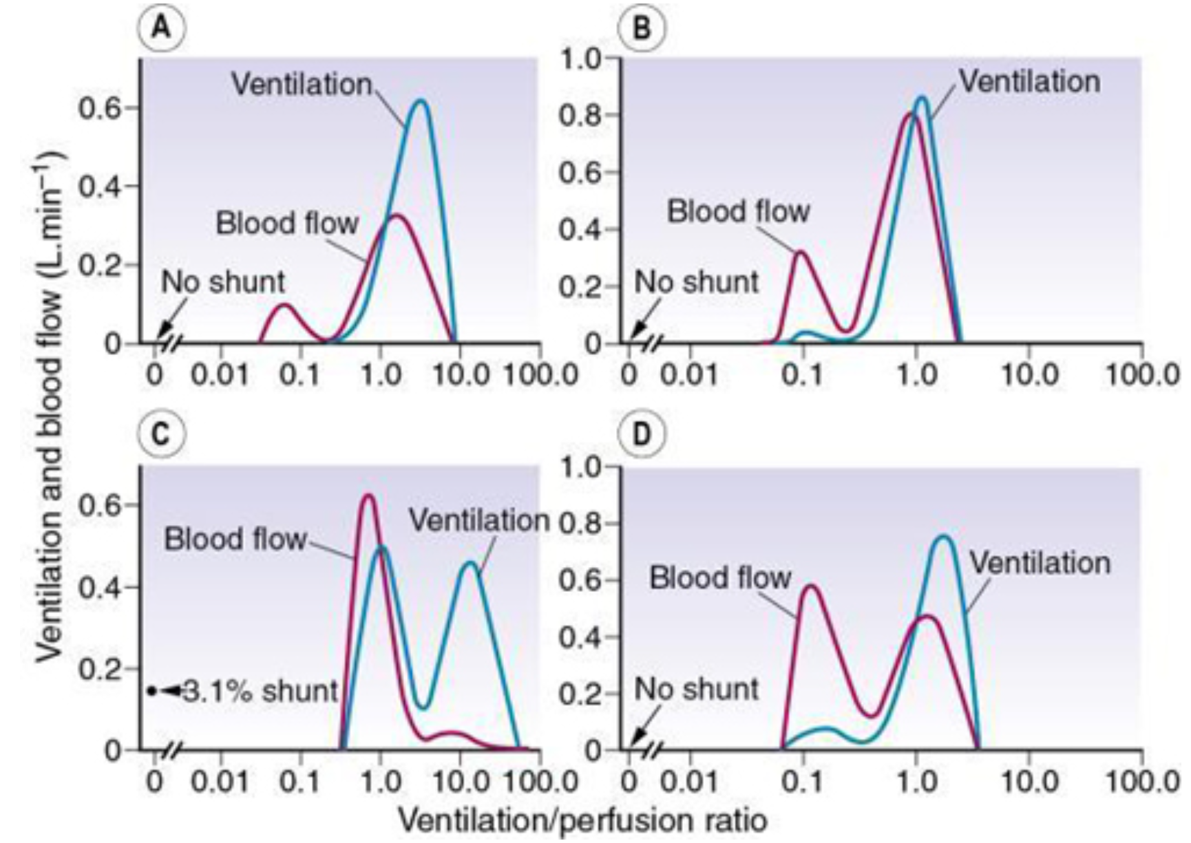
Examples of abnormal patterns of maldistribution of ventilation and perfusion, to be compared with the normal curves in Figure 7.8. (A) Chronic obstructive pulmonary disease, the blood flow to units of very low ventilation/perfusion ratio would cause arterial hypoxaemia and simulate a shunt. (B) Asthma, with a more pronounced bimodal distribution of blood flow than the patient shown in (A). (C) Bimodal distribution of ventilation seen in a 60-year-old patient with chronic obstructive pulmonary disease, predominantly emphysema. A similar pattern is seen after pulmonary embolism. (D) Pronounced bimodal distribution of perfusion after a bronchodilator was administered to the patient shown in (B). Source: (From West JB. Ventilation: Blood Flow and Gas Exchange. Oxford: Blackwell Scientific; 1990.With permission of the author and publishers.)
![]() Pink puffers and blue bloaters.pptx
Pink puffers and blue bloaters.pptx